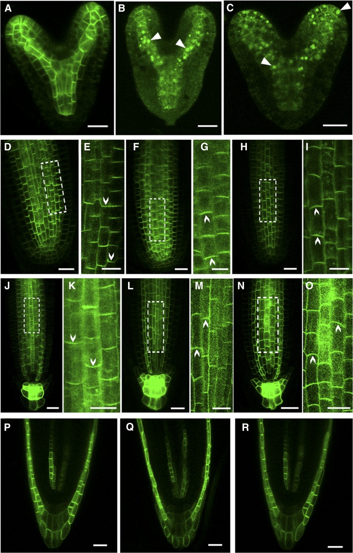
Figure 9.
ROP3 Is Required for the Polar Localization of PINs.
(A) to (C) PIN1pro:PIN1:GFP localization in embryos of the wild type (A), RPS5Apro:DN-rop3 (B), and rop3 (C). Arrowheads indicate intracellular aggregates of PIN1 protein.
(D) to (I) PIN1pro:PIN1:GFP localization in roots of 4-d-old wild-type ([D] and [E]), 35Spro:DN-rop3 ([F] and [G]), and rop3 ([H] and [I]) seedling. Regions in white boxes (D), (F), and (H) are confocal scans with high magnifications shown in (E), (G), and (I). Arrowheads indicate the direction of PIN1 polarity.
(J) to (O) PIN3 pro:PIN3:GFP localization in roots of 4-d-old wild-type ([J] and [K]), 35Spro:DN-rop3 ([L] and [M]), and rop3 ([N] and [O]) seedling. Regions in white boxes ([K], [M], and [O]) are confocal scans at high magnification. Arrowheads indicate the direction of PIN3 polarity.
(P) to (R) AUX1pro:AUX1:YFP localization in roots of 4-d-old wild-type (P), 35Spro:DN-rop3 (Q), and rop3 (R) seedling.
Bars = 20 μm.
[See online article for color version of this figure.]

