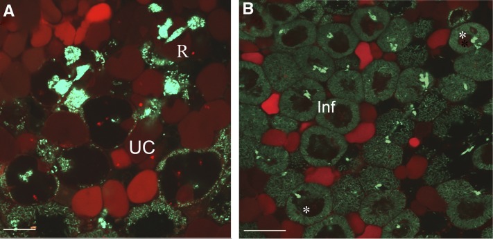
Figure 3.
NR Staining to Determine Vacuolar pH in Nodule Cells.
(A) Infection zone.
(B) Fixation zone.
Confocal images display the red color of the acidotrophic dye showing acid compartments–vacuoles. Symbiosomes were counterstained by Sytox Green. inf, infected cells; R, bacteria release; UC uninfected cells. Asterisks indicate dead bacteria stained by NR inside the vacuole lumen. Bar in (A) = 20 µm; bar in (B) = 50 µm.

