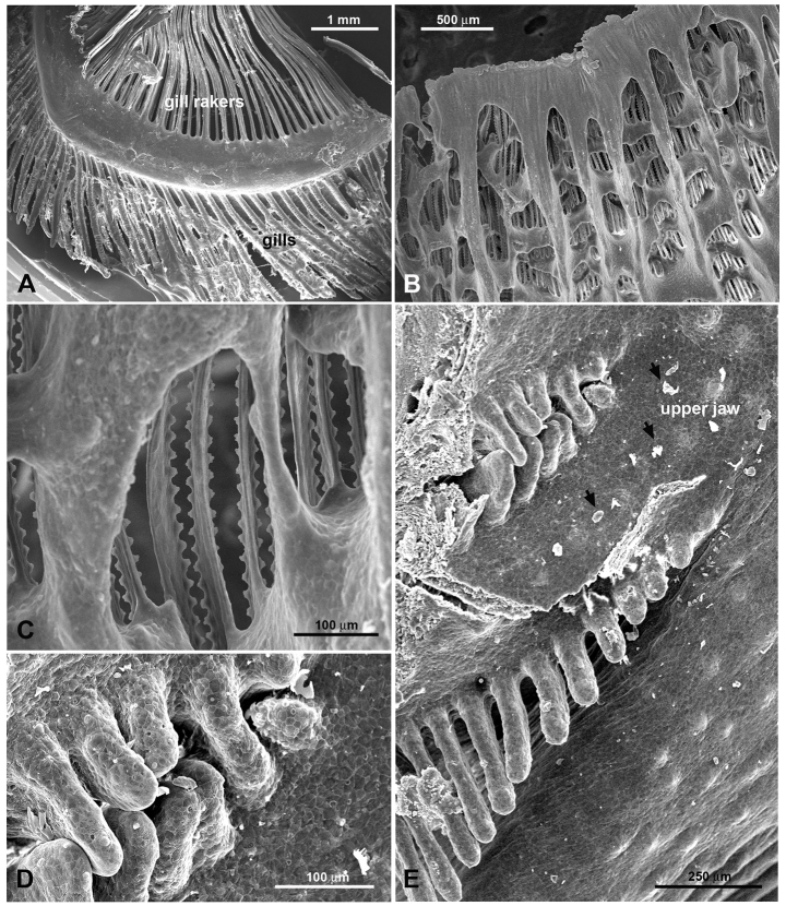
Fig. 2.
Scanning electron microscope images of gills and gill rakers in H. nobilis and H. molitrix. (A) Branchial arch with gills and gill rakers in H. nobilis. The gill rakers are free. (B) Gill rakers in H. molitrix. The gill rakers are widely fused and build a sieve-like structure. (C) Higher magnification of fused gill rakers in H. molitrix. (D,E) Entrance to the EO tubes at the posterior end of the upper jaw of H. nobilis. Small food particles (arrows) litter the epithelium.

