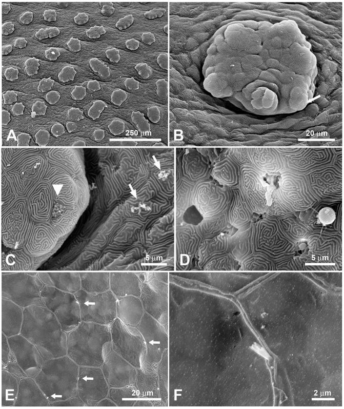
Fig. 3.
Scanning electron microscope images of the outside of the epibranchial organ in H. molitrix. (A) The outside epithelium of the epibranchial organ is densely covered with small protrusions. (B) Each protrusion itself has further protrusions. Taste buds are inserted in these protrusions (arrow). (C) Higher magnification of one of these small protrusions showing the taste pore of a taste bud (arrowhead). The surface of the epithelium is littered with small food particles (arrows). (D) Abundant mucus cells are present throughout the epithelium. This image shows the various stages of mucus cells: discharged (left), discharging (middle) and discharged droplets of mucus (right). Almost all epithelial cells of the epibranchial organ show the ‘finger-print’ pattern typical for fishes. (E) Numerous solitary chemosensory cells (arrows) are scattered between the epithelial cells. (F) Higher magnification of a solitary chemosensory cell. The apex of these cells varies between oligovillous as seen here and monovillous.

