Fig. 5.
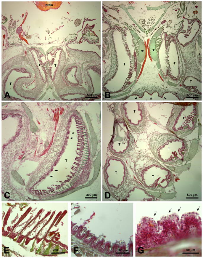
Histological staining with nuclear red/light green/orange of 14 μm cryosections of the EO. Cartilage and bone were stained green, the brain, nerves and nerve fiber bundles were stained orange, and epithelia including taste buds were stained purple. These stainings were done for a general overview on sections adjacent to the sections used for immunohistochemistry. Images A, B and C show H. molitrix; images D, E, F and G show H. nobilis. A, B and C depict cross-sections through the epibranchial organ and its tubes (T). Image B shows the supporting cartilaginous structures (*) and the ridges at the outside of the epibranchial organ (arrowheads). (D) Horizontal section: the tubes are lined with epithelium that contains taste buds (arrows in C). In some areas modified gill rakers face the areas with taste buds. (E) Higher magnification of the modified gill rakers. (F) Some areas of the tubes contain small flaps as shown in Fig. 4C. (G) Higher magnification of the small flaps, which are lined with abundant mucus cells (arrows).

