Fig. 6.
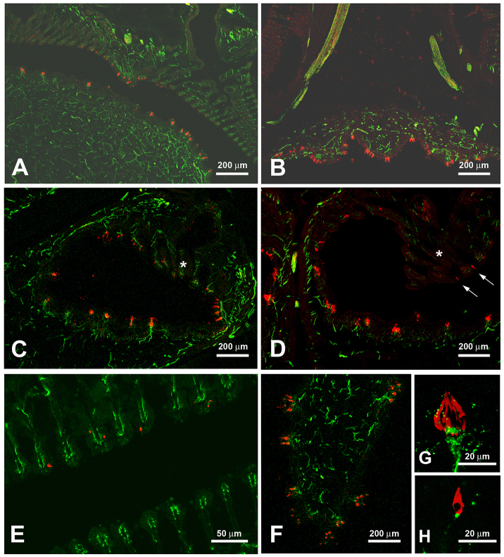
Histology of the epibranchial organ. Sections adjacent to those of Fig. 5 were treated with antibodies against calretinin (red), a marker for taste buds and solitary chemosensory cells, and acetylated tubulin (green), a marker for nerve fibers. Images A, B, D and E show H. molitrix, images C, F, G and H show H. nobilis. (A) As seen in Fig. 5, parts of the tubes are lined with epithelium containing taste buds whereas taste buds in other areas are scarce. (B) The epithelium outside the EO has ridges that contain taste buds (cf. Fig. 5B). (C) Taste buds innervated by small tubulin-positive nerve fibers lie opposite modified gill rakers (*), which usually do not have taste buds. (D) The modified gill rakers (*) contain few solitary chemosensory cells (arrow). (E) Higher magnification of the modified gill rakers. The red dots depict a few calretinin-positive solitary chemosensory cells. (F) The ridges on the outside of the EO have small protrusions that contain several taste buds. (G) Higher magnification of a taste bud (red) contacted by small nerve fibers (green). (H) Higher magnification of a solitary chemosensory cell also contacted by small nerve fibers.

