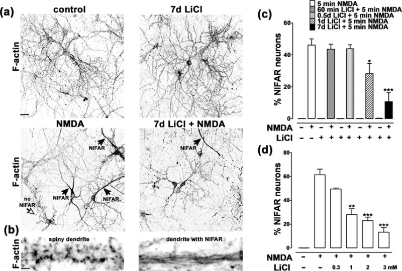
Figure 1. Lithium prevents NMDA-induced F-actin reorganization.

(a) Cultured hippocampal neurons incubated in the absence (top left) or presence (bottom left) of NMDA (5min; 50 μM), with (right) or without (left) LiCl and then fixed and stained for F-actin using Alexa568-phalloidin. The black arrows indicate neurons that have undergone NMDA-induced F-actin reorganization (NIFAR), while the open arrow indicates a no-NIFAR neuron. We observed that NIFAR occurs only in the presence of NMDA and that it is significantly attenuated by preincubation with LiCl. Scale bar: 50 μm. (b) Higher magnification images from dendritic regions of a control (left) and a NIFAR neuron (right). Scale bar: 3 μm.
(c) Time course of effect of LiCl preincubation on prevention of NIFAR. Hippocampal neurons were incubated with 3mM LiCl for the indicated times prior to addition of 50 μM NMDA for 5 min. Data are expressed as the percentage of neurons exhibiting NIFAR and graphed as mean ± SEM (*p<0.05, *** p<0.001 one-way ANOVA, followed by Bonferroni post hoc test vs NMDA +; number of coverslips: 6 per condition; number of neurons counted per coverslip = 400–800). NIFAR was absent in untreated cultures or cultures treated with lithium alone.
(d) Dose-dependence of the effect of LiCl preincubation on prevention of NIFAR. Hippocampal neurons were pre-incubated for 7 days with the indicated concentrations of LiCl prior to addition of 50 μM NMDA for 5 min. Data are expressed as percentage of neurons exhibiting NIFAR, and graphed as mean ± SEM. (*p<0.05, *** p<0.001 one-way ANOVA, followed by Bonferroni post hoc test vs NMDA +; number of coverslips: 6 for each condition; number of neurons counted per coverslip = 400–800). NIFAR was absent in untreated cultures.
