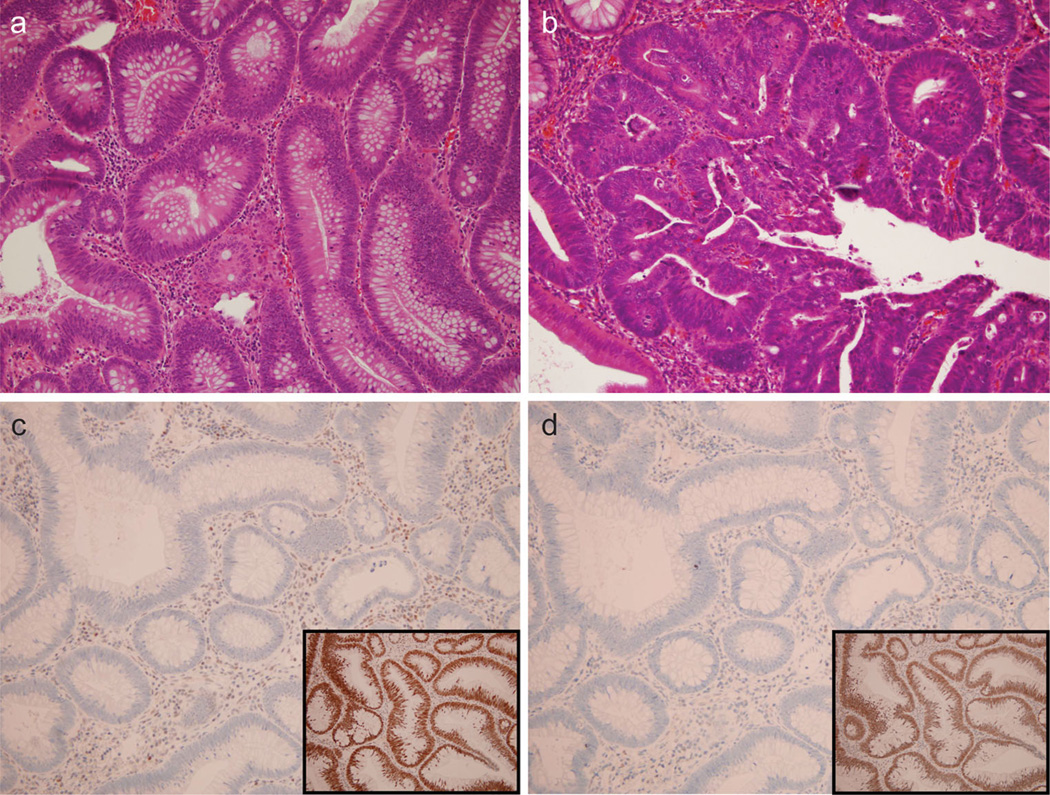
Fig. 1.
Histologic features of advanced adenomas and MMR immu-nohistochemistry. a Typical adenoma (2009 magnification, H&E stain). b Adenoma in another patient with high-grade dysplasia displaying loss of nuclear polarity and cribriform gland architecture (400× magnification, H&E stain). c Loss of MLH1 nuclear positivity (200× magnification, IHC) in the 19-year-old female subject from this study. Note the positive control example of retained MLH1 (inset) in a different subject. d Loss of PMS2 nuclear positivity in the same 19-year-old female subject as panel C (200× magnification, IHC). Note the positive control example of retained PMS2 in a different subject (inset)

