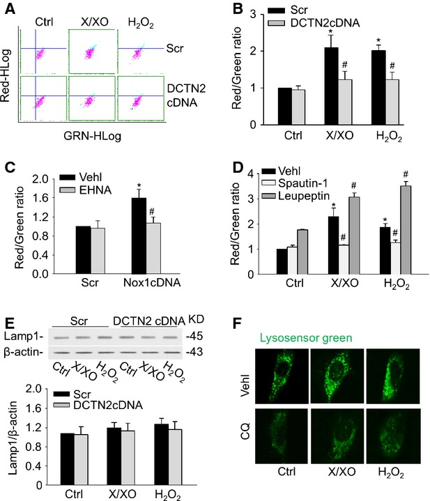
Figure 4.

Inhibition of dynein activity decreased APLs formation. Mouse CAMs were stained with acridine orange for 17 min. Representative dot plots of flow cytometry (A) and summarized red-to-green fluorescence ratio analysis showing APLs formation in CAMs with DCTN2 cDNA (B) or EHNA (C). (D) Summarized red-to-green fluorescence ratio analysis showing APLs formation in CAMs with spautin-1 (10 μM) and leupeptin (0.25 mM). (E) Representative Western blot documents showing the expression of Lamp-1 from CAMs. (F) CAMs were treated with chloroquine (CQ, 100 μM) for 30 min. or left untreated. They were then stained with LysoSensor Green DND-189 and analysed by fluorescence microscopy (n = 6 for all panels). *P < 0.05 versus Scramble; #P < 0.05 versus CAMs with X/XO or H2O2 or Nox1 cDNA transfection alone.
