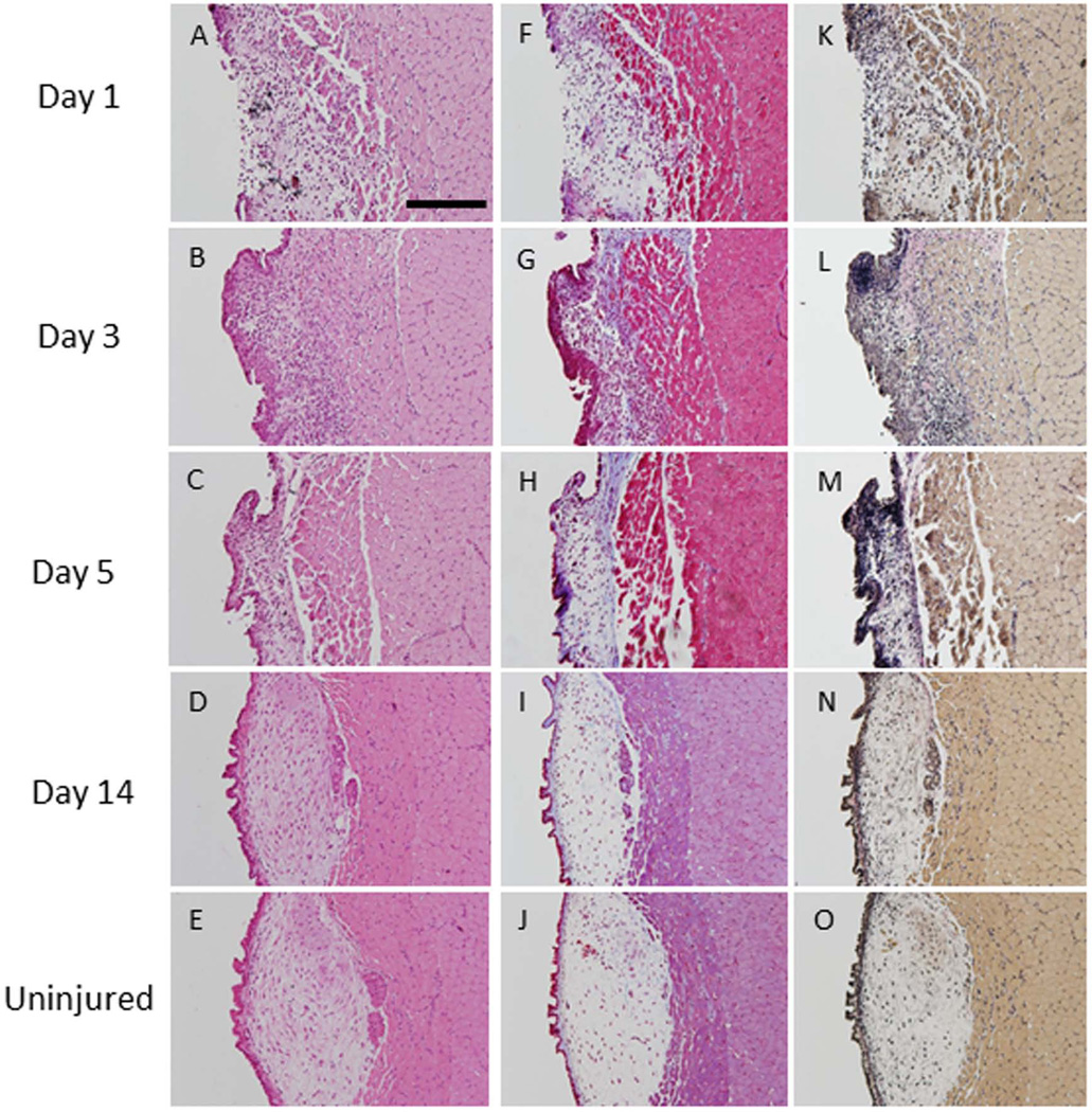
Figure 2. Subepithelial injury.
Hematoxylin and eosin staining at three time points post-injury (A–D) and in an uninjured vocal fold (E). Trichrome staining shows increased collagen density (blue) up to 5 days post-injury (F–H). Collagen deposition at day 14 (I) were indistinguishable from levels observed in uninjured tissue (J). Verhoff–van Gieson (VVG) staining showed increased elastin deposition (black) in the injured lamina propria up to 5 days post-injury (K-M) compared to uninjured vocal folds (O). At day 14 (N), elastin deposition was at uninjured levels (O) (200µm scale bar; original magnification 20×).

