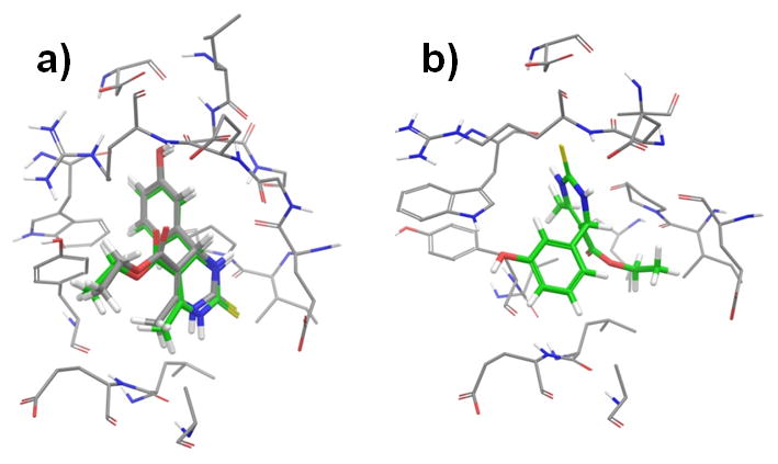
Figure 4.

Comparison of the IFD generated docked models at the monastrol-binding site. a) Docked model-a (green-colored carbon) well reproduced the binding mode of the crystal structure (grey-colored carbon). b) Docked model-b bound very differently from model-a. The monastrol molecules were shown in solid sticks. Eg5 residues within 4 Å of the ligands were shown in thin tubes.
