
Fig. 1.
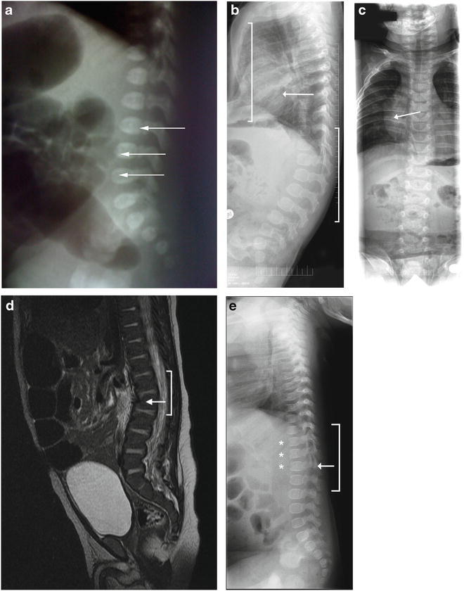
Skeletal abnormalities in infants with MPS II. (a) Skeletal X-ray image of Patient 1 at 1 day of age showing L3-L5 abnormality (arrows). b and c) Skeletal X-ray images of Patient 2 at 6 months of age showing pectus excavatum (panel b , left bracket), mild lumbar kyphosis (panel b , right bracket), and mild broadening of the ribs (panels b and c , arrows). (d) A computed tomography scan of Patient 4’s abdomen performed at 8 weeks of age showing upper lumbar kyphosis (bracket) with mild inferior beaking at L2 (arrow). (e) Skeletal X-ray images of Patient 8 at 5.5 months of age showing a thoracolumbar gibbus (bracket) with the apex at L2 (arrows) and beaked vertebrae (stars)