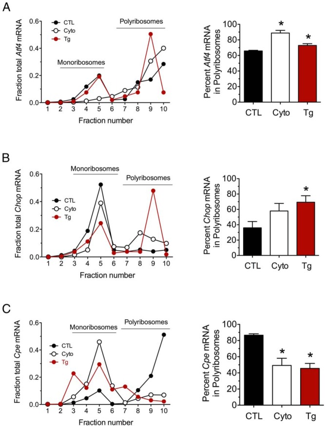
Figure 3.
Effect of proinflammatory cytokines and thapsigargin on ribosomal occupancy of Atf4, Chop, and Cpe. MIN6 β cells were untreated (CTL), treated with proinflammatory cytokines (Cyto) for 24 hours, or treated with thapsigargin (Tg) for 4 hours and then were subjected to PRP analysis with fractionation of the sedimentation gradient. A, Analysis of Atf4 mRNA in PRP fractions. B, Analysis of Chop mRNA in PRP fractions. C, Analysis of Cpe mRNA in PRP fractions. Representative data are shown on the left in A, B, and C, and quantitative data on the percentage of total mRNA residing in polyribosomes (fractions 6–10) are shown on the right. Data represent means ± SEM (n = 3–5). *, P < .05 compared with the control.

