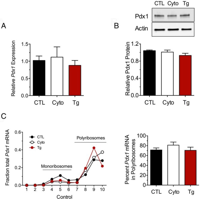
Figure 5.
Pdx1 retains ribosomal occupancy in the setting of UPR activation. A, MIN6 β-cells were untreated (CTL), treated with cytokines (Cyto) for 24 hours, or treated with thapsigargin (Tg) for 4 hours and then were subjected to qRT-PCR, immunoblot analysis, or PRP analysis with fractionation of the sedimentation gradient. A, qRT-PCR analysis in whole-cell extracts. B, Immunoblot analysis (top panel) with corresponding quantitation (n = 3–5, lower panel). C, qRT-PCR analysis of Pdx1 mRNA in PRP fractions (left panel) and corresponding quantitation of the percentage of total mRNA in polyribosomes. Data represent means ± SEM (n = 3–6).

