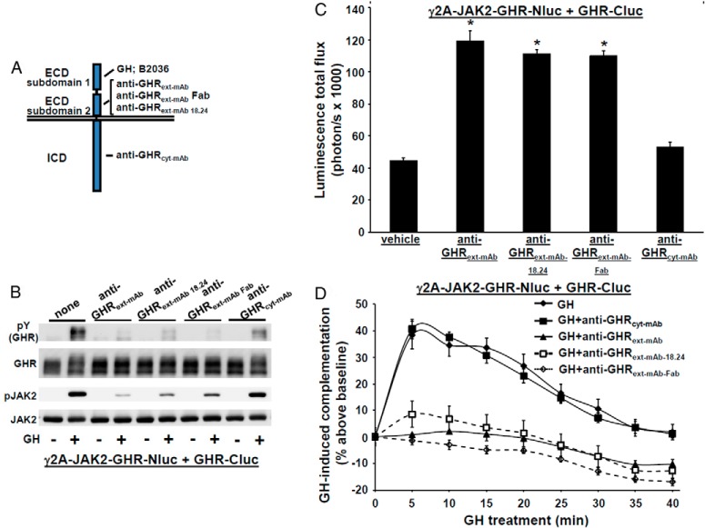
Figure 3.
Effect of antagonistic GHR antibodies on GHR-GHR complementation. A, Diagram of regions of GHR that interact with each monoclonal antibody (or Fab) used in this study, GH, or the GH antagonist, B2036. The regions indicated are subdomains 1 and 2 of the ECD and the ICD. B, GHR-specific monoclonal antibodies or a Fab fragment specifically inhibit GH-induced phosphorylation. γ2A-JAK2-GHR-Nluc cells transiently expressing GHR-Cluc cells were serum starved, preincubated with anti-GHRext-mAb (40 μg/mL), anti-GHRext-mAb-18.24 (40 μg/mL), anti-GHRext-mAb-Fab (13.3 μg/mL), or anti-GHRcyt-mAb (40 μg/mL) (a control mAb against the GHR ICD) for 30 minutes, and then treated ±GH (500 ng/mL, 10 min). Detergent cell extracts were resolved by SDS-PAGE and sequentially immunoblotted to detect phosphorylated GHR-luciferase chimeras (with anti-pY) and total GHR-luciferase chimeras and phosphorylated and total JAK2. The blot shown is representative of three such experiments. C, Treatment with GHR-specific monoclonal antibodies or a Fab fragment specifically augments basal (non-GH dependent) GHR-GHR complementation. γ2A-JAK2-GHR-Nluc cells transiently expressing GHR-Cluc were treated with vehicle, anti-GHRext-mAb (40 μg/mL), anti-GHRext-mAb-18.24 (40 μg/mL), anti-GHRext-mAb-Fab (13.3 μg/mL), or anti-GHRcyt-mAb (40 μg/mL, a control mAb against the GHR ICD) for 30 minutes, after which bioluminescence was determined. Data are displayed graphically as mean ± SE total flux (photons per second × 1000). *, P < .05 compared with vehicle treatment. The figure shown is representative of three such experiments. D, GHR-specific monoclonal antibodies or Fab fragment specifically inhibit GH-induced changes in GHR-GHR complementation. γ2A-JAK2-GHR-Nluc cells transiently expressing GHR-Cluc were treated with GH (500 ng/mL) or GH plus the indicated antibodies (40 μg/mL) or Fab fragment (13.3 μg/mL) after basal bioluminescence was determined. Bioluminescence was measured serially thereafter over 40 minutes. Data are expressed as mean ± SE of GH-induced signal as a percentage above baseline signal (n = 3 per condition). Baseline signal is normalized for each condition. For GH vs GH+anti-GHRext-mAb/anti-GHRext-mAb 18.24/anti-GHRext-mAb Fab, the value was P < .05 at each time point; for GH vs GH+anti-GHRcyt-mAb, the value was P > .05 at each time point. The figure shown is representative of three such experiments.

