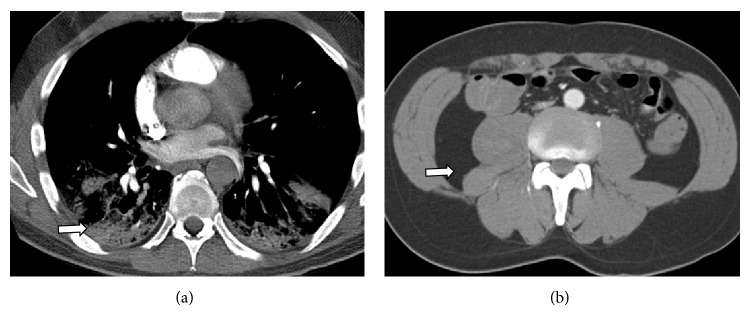
Figure 1.

(a) CT chest on admission. Axial contrast enhanced images through the chest in the pulmonary phase demonstrate a normal main pulmonary artery trunk and normal segmental pulmonary arterial branches extending to the superior segment of the right upper lobe. Lung windows reveal lower lobe consolidation (see arrow). (b) Normal appearance of the retroperitoneum at that time (see arrow).
