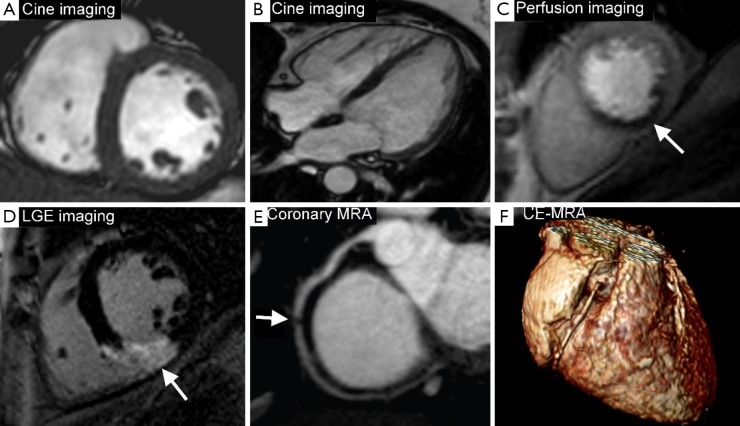
Figure 1.
CMR techniques. Panels A and B are cine images (short-axis and 4-chamber views respectively) which give anatomical and functional information. Panel C is a stress perfusion CMR showing an inferior perfusion defect (white arrow) in the mid-ventricular slice. Panel D is LGE imaging showing inferior wall transmural myocardial infarction (white arrow). Panel E shows coronary MRA demonstrating a mid right coronary artery stenosis. Panel F is an example of 3D volume rendered contrast enhanced MRA showing the right coronary artery. CMR, cardiovascular magnetic resonance; LGE, late gadolinium enhancement; MRA, magnetic resonance angiography; 3D, three-dimensional.

