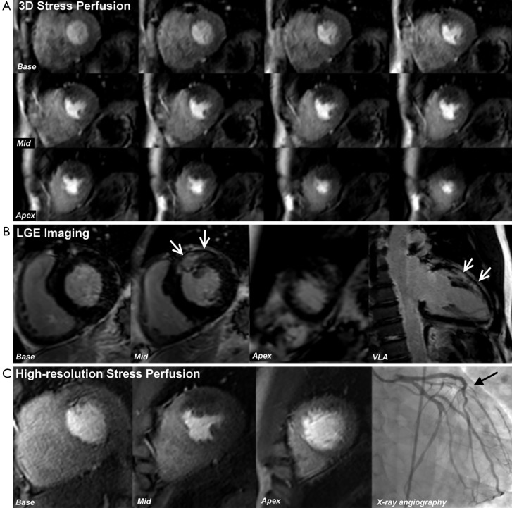
Figure 5.
3D whole heart perfusion CMR. A 45-year-old man with previous PCI to the LAD presented with significant angina. (A) shows 3D perfusion CMR (12 slices) at stress; (B) shows LGE imaging and (C) shows high-resolution (1.1 mm in-plane) stress perfusion CMR—all performed at 3.0 T. 3D perfusion CMR shows stress-induced hypo-perfusion throughout the anterior wall from base to apex—i.e., well beyond the area of scar seen in the mid-anterior wall on LGE imaging. This example shows the benefit of whole-heart coverage with the 3D acquisition, as the 3-slice high-resolution techniques did not demonstrate any significant ischemia beyond the established scar in the mid-ventricle. X-ray angiography confirmed a sub-total occlusion of a large diagonal branch, accounting for the anterior ischemia (black arrow) (Reprinted with permission from: Motwani et al. Circ Cardiovasc Imaging 2013;6:339-48) (29). 3D, three-dimensional; CMR, cardiovascular magnetic resonance; VLA, vertical long axis.

