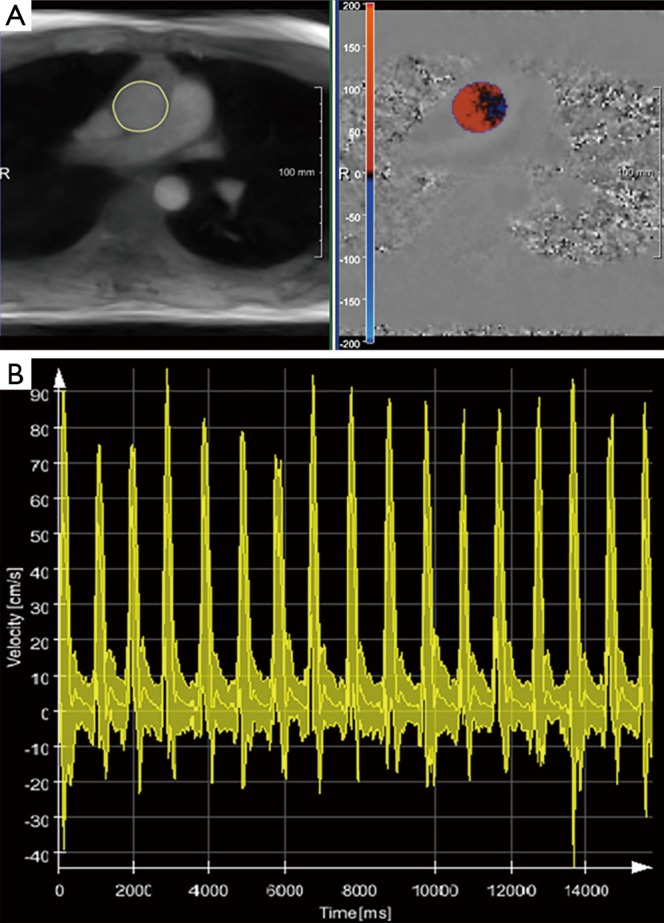
Figure 12.

Real-time phase-contrast flow MRI (3 T, 40 ms): Quantitative evaluations for the ascending aorta of a healthy subject using CAIPI software. (A) End-systolic magnitude image and phase-contrast map with segmented ascending aorta and color-coded flow velocities, respectively; (B) the overlay of peak flow velocity, mean velocity averaged over the lumen of the aorta and minimum velocity as a function of time for 17 consecutive heartbeats demonstrates the influence of respiration. MRI, magnetic resonance imaging.
