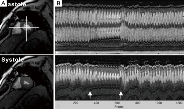
Figure 9.

Real-time cardiac MRI with T1 contrast (3 T, 33 ms) of a patient with diastolic dysfunction during Valsalva maneuver, i.e., increased intrathoracic pressure. (A) End-diastolic and end-systolic images from a corresponding movie (see Figure 10); (B) temporal intensity profiles (40 s, top = horizontal reference, bottom = vertical reference) demonstrate reduced systolic and diastolic myocardial movements as well as alterations of wall thickening during Valsalva maneuver (arrows). MRI, magnetic resonance imaging.
