
Figure 10.
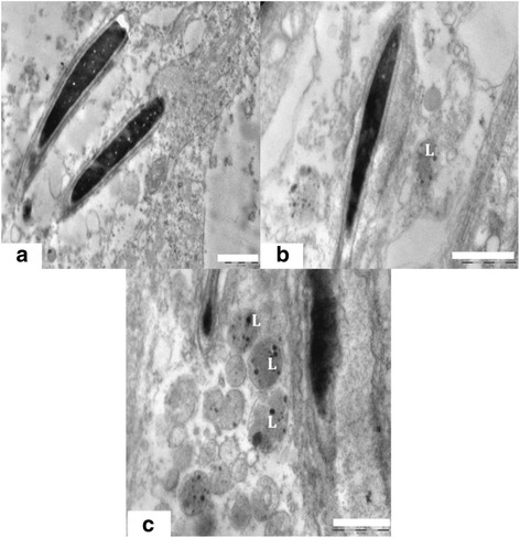
TEM of spermatids & lysosomal bodies. (a) Ultra structure of a normal spermatid with intact head, acrosomes and membrane. Scale bar =1 μm, (b) Presence of lysosomal bodies containing nanoparticles near the elongating spermatids and deformed acrosomal membrane in experimental group. Scale bar =1 μm and (c) Sertoli cell cytoplasm crowded with lysosomal bodies containing nanoparticles and abnormal spermatids. Scale bar =1 μm.