
Figure 11.
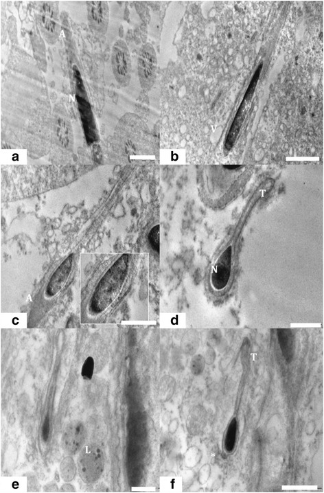
TEM of elongated spermatid. (a) Ultra structure of the testis of rat from control group 1 showed intact acrosome (A), nucleus (N) and other cellular architecture of elongated spermatid. Scale bar =1 μm, (b) Experimental group 2 treated with Nano particle showed severely damaged ultra structure of elongated spermatid with high vacuolation (V) and electron dense bodies. Scale bar =2 μm, (c) Elongated spermatid from treated group 2 having membrane abnormalities surrounded by degenerated sertoli cell cytoplasm. Scale bar =1 μm, (d) Elongated spermatid from control group 1 with intact head and tail (T). Scale bar =0.5 μm, (e) Elongated spermatid from experimental group 2 deformed tail and head with increased sub acrosomal space. nanoparticle entrapped in lysosmal bodies (L) near the elongated spermatid was also noted. Scale bar =0.5 μm and (f) Elongated spermatid containing deformed acrosomal membrane (*) and tail (T) were commonly observed. Scale bar =1 μm.