
Figure 7.
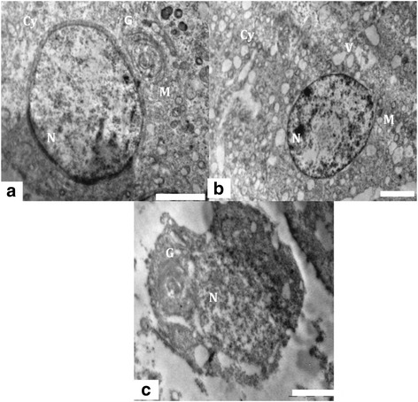
TEM of Spermatid. (a) Ultra structure of the testis of control animals, showing a round spermatid having distinct nucleus (N), mitochondria (M) and golgi vesicle (G). Scale bar =2 μm, (b) Testis of experimental animal showing degenerated cytoplasm (Cy), vacuoles (V) and collapsed mitochondria (M). Scale bar =2 μm and (c) Ultra structure of apoptotic round spermatid showing collapsed golgi vesicles (G) and nucleus (N) from experimental group. Scale bar =2 μm.