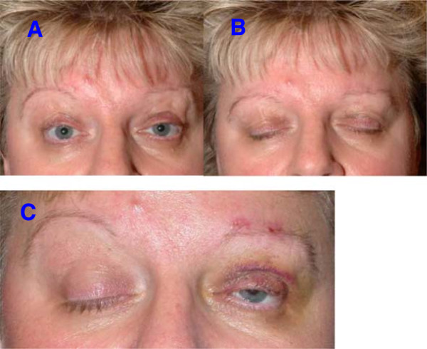
Figure 3.

Patient after bilateral frontalis suspension surgery. The eyes can be opened (A) and closed (B) without difficulty. The patient’s BoNT treatment was continued. Insert C shows the situation immediately after the unilateral operation of the left eye, identifiable by the small hematoma. One can clearly see that the apraxia persists in the non-operated right eye and that eye opening is not possible in spite of innervation of the frontalis muscle. The position of the eyebrow is higher and the upper eyelid is completely closed. After the suspension surgery, the left, operated eye is already partly opened with only a low-intensity innervation. This is consistent with a lateralized control of the apraxia.
