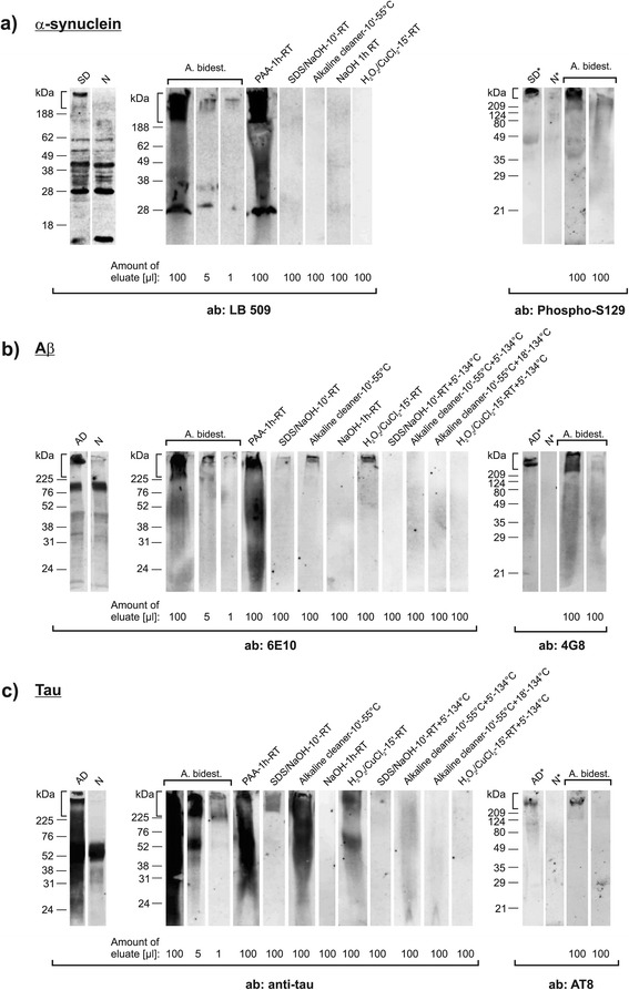
Figure 1.

In vitro carrier assay for testing the depletion of aggregated human α-synuclein, amyloid-β and tau. Western blot detection of aggregated human α-synuclein (a), Aβ (b), and tau (c) by the indicated antibodies (Table 1) in protein eluates from steel wire grids that had been contaminated with 20% (w/v) brain tissue homogenates (BTH) from donors with SD (a) or AD (b and c). Lanes “SD” (a), “AD” (b and c) and “N” (a-c) (“SD*” [a], “AD*” [b and c] and “N*” [a-c]) represent 5 μl (or 20 μl in lanes marked with an asterisk) 20% (w/v) BTH from SD- and AD patients and control donors (without SD or AD), respectively. The identity of α-synuclein, Aβ, and tau aggregates in BTH and on steel wire grids washed with bi-distilled water was confirmed by two different antibodies each (most right lanes in a-c represent steel wire grids that had been contaminated with BTH from control donors). For testing the presence and depletion of aggregated α-synuclein, Aβ, and tau contaminated wires were processed by I) washing with bi-distilled water (a-c, lanes “A. bidest.”), or exposure to II) 0.25% (v/v) peracetic acid for 1 hour at room temperature (RT) (a-c, lanes “PAA-1 h-RT”), III) a mixture of 0.2% (w/v) SDS and 0.3% (w/v) NaOH (pH 12.7 - 12.9, non-adjusted) for 10 min. at RT (a-c, lanes “SDS/NaOH-10'-RT”), IV) an alkaline cleaner (0.5%, pH 11.6 - 12.0 [non-adjusted]) [13] for 10 min. at 55°C (a-c, lanes “Alkaline cleaner-10'-55°C”), V) 1 M NaOH (pH 13.5 - 13.8, non-adjusted) for 1 h at RT (a-c, lanes “NaOH-1 h-RT”), VI) a solution of 7.5% (w/v) H2O2 containing Cu2+ ions [14] (prepared in 100 mM NaCO3 [pH 9.5, adjusted] by adding CuCl2 to a final concentration of 500 μM) for 15 min. at RT (a-c, lanes “ H2O2/CuCl2-15′-RT”), VII) treatment as in III) with subsequent PVSS for 5 min. at 134°C (b and c, lanes “SDS/NaOH-10′-RT + 5′-134°C”), VIII) treatment as in IV) with subsequent PVSS for 5 min. (b and c, lanes “Alkaline cleaner-10′-55°C + 5′-134°C”) or 18 min. (b and c, lanes “Alkaline cleaner-10′-55°C + 18′-134°C”), or IX) treatment as in VI) with subsequent PVSS for 5 min. (b and c, lanes “H2O2/CuCl2-15′- RT + 5′-134°C”). PVSS was always performed at 3 bar. ab: antibody. Vertical brackets indicate insoluble α-synuclein-, Aβ- and tau aggregates retained in stacking gels.
