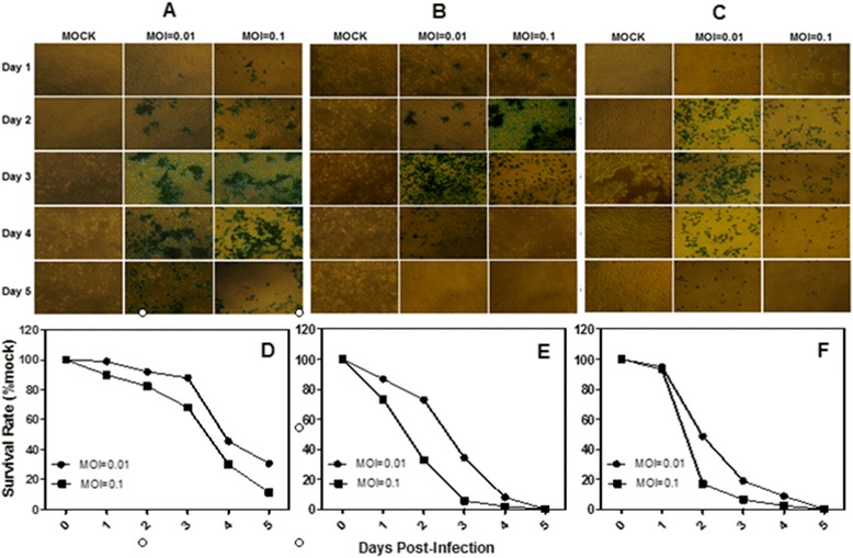
Figure 2.

The cytotoxicity of two HCC cell lines and one human hepatic immortalized cell line. A, X-gal staining of BEL-7404 cells infected with G47Δ. Monolayers of BEL-7404 cells in 6-well dishes were infected with G47Δ or control vehicle and incubated with DMEM/1% heat-inactivated FBS at 37°C. On each of the 5 days following infection, the cells were stained with X-gal solution. The infected cells express Lac Z and stained blue. B, X-gal staining of SMMC-7721 cells infected with G47Δ. C, X-gal staining of HL-7702 cells infected with G47Δ. D, E and F, Monolayers of BEL-7404, SMMC-7721 and HL-7702 cells in 6-well dishes were infected with G47Δ with MOI = 0.01 or MOI = 0.1, incubated in DMEM/1% heat-inactivated FBS at 37°C, and counted using a Coulter Counter on the days indicated. The average numbers of cells from duplicate wells are plotted as percentages of the mock wells.
