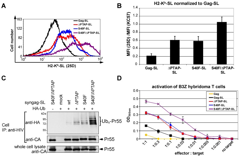
Figure 7.
The S40F mutation induces an enhanced MHC-I antigen presentation of Gag derived epitopes. (A) HeLa-Kb cells were transfected with syngag expression constructs coding for Gag-SL, ΔPTAP-SL, S40F-SL and S40F/ΔPTAP-SL. H2-Kb-SL complexes presented on the surface of Gag-positive cells were quantified by flow cytometry using 25D1.16-APC. A representative histogram plot is shown; (B) Quantification of three independent experiments. The mean fluorescence intensity (MFI) of the 25D1.16 staining was normalized to the MFI of the intracellular anti-Gag staining obtained after permeabilization of the plasma membrane. Bars represent mean values ± SD; (C) Analysis of the Gag ubiquitination of the corresponding constructs. Gag was recovered from whole cell lysates by immunoprecipitation using anti-HIV antibodies, and ubiquitinated species were detected by anti-HA staining; (D) Activation of B3Z hybridoma T cells was assessed by a colorimetric β-galactosidase assay after overnight cocultivation with HeLa-Kb cells expressing Gag wt, Gag-SL wt, ΔPTAP-SL, S40F-SL or S40F/ΔPTAP-SL in various effector-to-target ratios.

