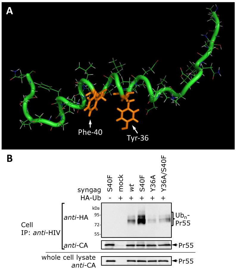
Figure 9.
NMR structure of p6(23-52)S40F confirms formation of a new hydrophobic domain. (A) Structure of sp6(23-52)S40F determined by NMR spectroscopy. The residues Y36 and F40, which are located at the same surface of the helix, forming a new hydrophobic domain within the C-terminal helix of p6, are labeled in orange. (B) HeLa cells were co-transfected with HA-tagged ubiquitin and the syngag expression plasmids directing the expression of wt Gag and mutants thereof. The isolation and detection of ubiquitinated Gag species from cell fractions were carried out as described in Figure 2B. The amount of Gag recovered by immune precipitation and Gag recovered from whole cell lysate was detected with CA specific antibodies.

