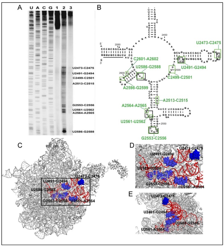
Figure 4.
(A) Sequencing gel showing the binding (road block) sites of 6AP (lane 2) and 6APi (lane 3) on the domain V rRNA identified by primer extension after UV-crosslinking sites. Lane 1 is the control without 6AP/6APi. (B) Binding sites of 6AP and the protein folding substrates in domain V rRNA. The green and the black boxes indicate 6AP and the protein binding sites, respectively. (A–B), The research was originally published in JBC [46]; Reproduced with permission. © The American Society for Biochemistry and Molecular Biology); (C) The position of the binding sites in the 50S subunit of E. coli ribosome. (PDB ID: 3UOS) The red highlighted portion represents residues 2435–2668 of domain V rRNA where most of the binding sites (marked with blue spheres and labeled with residue numbers) are located; (D) and (E) The enlarged view of the binding pocket (black box in C) from front (D) and side (E) view.

