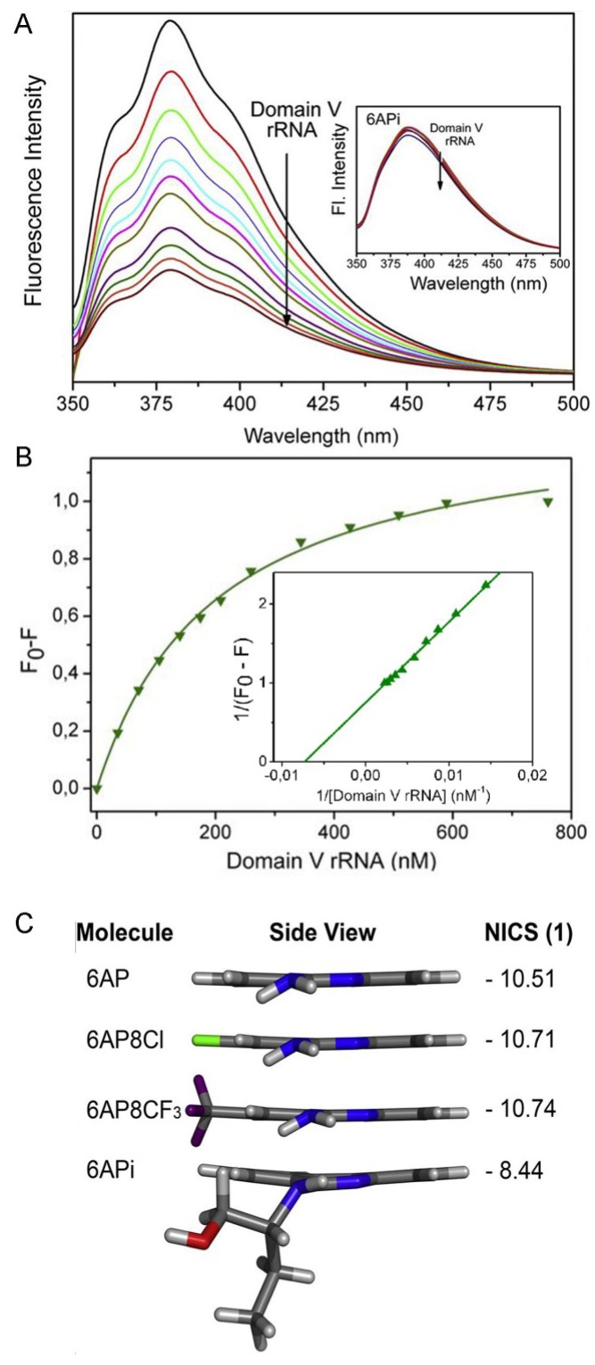
Figure 5.
(A) Quenching of 6AP fluorescence in the presence of domain V rRNA. Inset shows 6APi fluorescence, which does not quench by addition of domain V rRNA; (B) Saturation binding curve of 6AP with domain V rRNA. The double reciprocal plot of the binding curve is in the inset, the KD value is determined from the reciprocal of the X-intercept; (C) Lateral view of the 6AP derivatives and corresponding values of Nucleus independent chemical shift (NICS). The figure is reproduced with permission from [45]. Copyright © 2014 Elsevier Masson SAS. All rights reserved.

