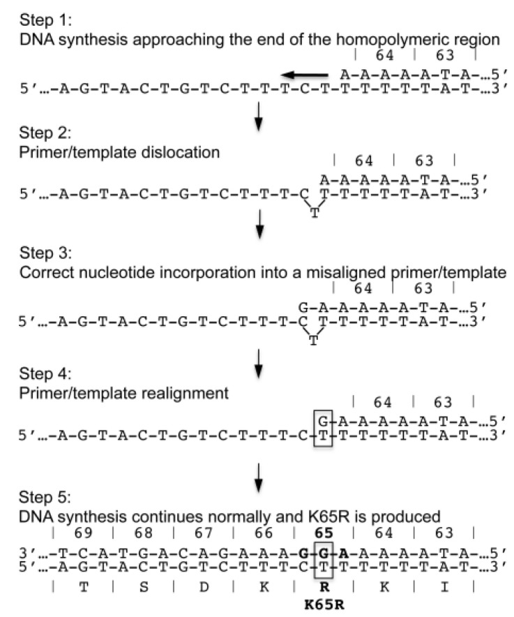
Figure 2.
Schematic of dislocation mutagenesis and the development of the K65R mutation in subtype C HIV-1. Step 1: DNA synthesis approaches the end of a homopolymeric nt stretch that ends precisely at the location of K65R development. Step 2: At the end of the sequence, the RT enzyme exhibits characteristic pausing of DNA synthesis. The template-strand folds onto itself and exposes a C in the folded-over template strand. Step 3: A dGTP nt correctly binds opposite the C base of the misaligned template strand as DNA synthesis continues. Step 4: The primer and template strands realign and the same C base becomes re-exposed on the now correctly aligned template strand. Step 5: A second dGTP becomes incorporated opposite the re-exposed C on the correctly aligned primer/template strands and DNA synthesis continues normally. This series of events yields the AAG-to-AGG change that is responsible for the more facilitated appearance of the K65R mutation in subtype C HIV-1. Reproduced from [55].

