Fig. 1.
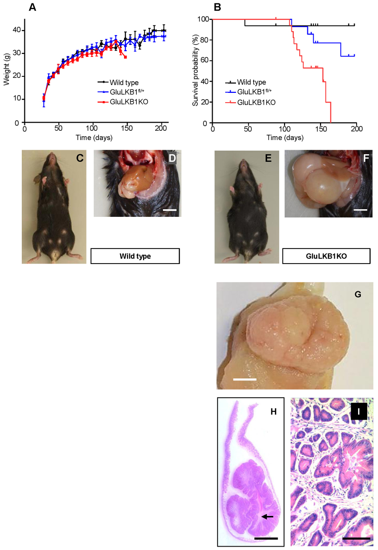
Decreased lifespan and development of gastro-duodenal polyps following Glu-Cre-dependent deletion of Lkb1. (A) Body weight changes in wild-type (n=10), GluLKB1fl/+ (n=11) and GluLKB1KO (n=13) mice. Analysis by one-way ANOVA did not demonstrate any significant difference between the three groups. (B) Kaplan-Meier survival curves for wild-type, GluLKB1fl/+ and GluLKB1KO mice. Vertical deflections on the graph represent censored data where the exact date of death was unknown owing to euthanasia of mice with ill health. GluLKB1KO mice (n=21; red), displayed significantly reduced lifespan and 100% of the cohort were deceased by day 164 (Log-rank Mantel-Cox test, P<0.001). GluLKB1fl/+ mice (blue; n=16) displayed a trend towards reduced longevity compared with the wild-type mice (black; n=16). By day 197, 93.7% of the wild-type mice had survived compared with 64.3% of the heterozygous mice and 0% of the homozygous knockout mice. Median survival for the GluLKB1KO mice was 153 days. (C) Representative wild-type mouse with normal phenotype. (D) Abdomen of a wild-type mouse displaying normal gastrointestinal contents. Scale bar: 5 mm. (E) Photograph of a representative GluLKB1KO mouse demonstrating a bloated appearance. (F) Photograph of the abdomen of a GluLKB1KO mouse displaying grossly distended stomach and duodenum due to the presence of a large gastro-duodenal polyp. Scale bar: 5 mm. (G) Gross macroscopy of a representative sessile polyp. (H) Haematoxylin-eosin (H&E)-stained section of a pedunculated polyp, demonstrating tubulovillous architecture with finger-like glands arising from the stalk and a core of arborizing smooth muscle (arrow). (I) Higher magnification of panel H. H&E staining with 10× magnification of the polyp. Scale bars: (G,H) 5 mm; (I) 100 μm.

