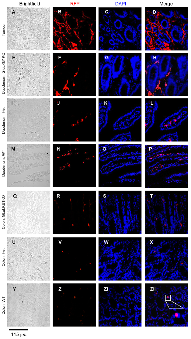
Fig. 3.
Fate of proglucagon-gene-expressing intestinal cells explored by lineage tracing. Immunohistochemistry of frozen sections using an anti-RFP antibody and confocal microscopy images of polyp, duodenum and colon from wild-type (‘WT’; M-P,Y-Zii), GluLKB1fl/+ (I-L,U-X) and GluLKB1KO (A-H,Q-T) mice. Brightfield, RFP, DAPI (nuclei localisation) and merged RFP/DAPI images are shown. In Zii, inset shows an enlarged image of an RFP-stained cell located within a crypt and with predicted enterocyte morphology.

