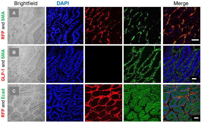
Fig. 4.
Immunohistological characterisation of RFP cells within polyps. Colocalisation experiments were performed on 7-μm frozen sections. (A) Anti-RFP (rabbit polyclonal) and SMA (mouse monoclonal), (B) GLP-1 (goat polyclonal) and SMA (rabbit polyclonal), and (C) RFP (rabbit polyclonal) and E-cadherin (mouse monoclonal). Brightfield, DAPI, red and green fluorescence, and the merged images are shown. RFP cells were found to colocalise with SMA; however, the SMA-labelled cells did not colocalise with either GLP-1 or E-cadherin. Scale bars: 50 μm.

