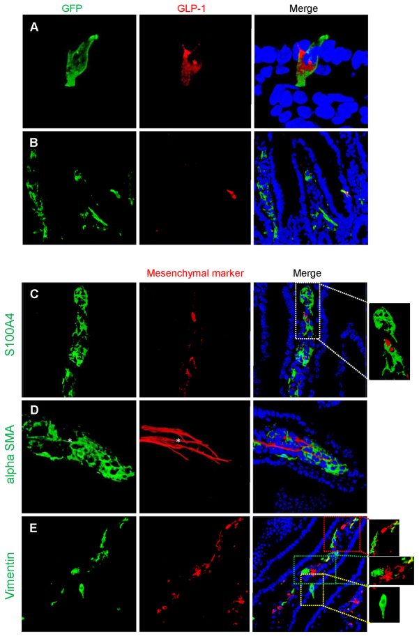
Fig. 5.
Expression of fluorescence in adult Glu-Cre mouse small intestine. (A) Photomicrographs demonstrating green fluorescence in a typical epithelial cell, resembling an enteroendocrine cell, and co-staining for proglucagon (GLP-1). The sequence in the panel shows the green fluorescence, proglucagon stained with a secondary Alexa-Fluor-555 and finally the merged image with Hoechst staining of the nuclei. (B) In certain cells, fluorescence is also demonstrated in mesenchymal cells within the villi; these cells do not co-stain for proglucagon. (C–E) Further characterisation of these GFP-positive but GLP-1-negative cells demonstrates that they do not co-stain with the fibroblast-specific marker S100A1 (C), but a proportion of cells co-stain with α-SMA (D) and others with vimentin (E). In this final panel (E), two areas of GFP and vimentin colocalisation are highlighted (red and green boxes) and a typical enteroendocrine cell is highlighted (yellow box). The asterisk in D indicates a GFP-positive cell representing a lacteal and co-staining with α-SMA.

