
Fig. 6.
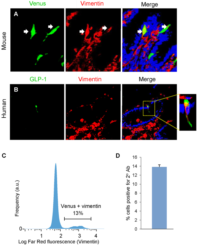
Characterisation of Venus proglucagon-expressing enteroendocrine cells. Adult mGLU124 upper small intestine was stained for GFP and vimentin, and a green and red secondary. (A) Some sporadic Venus cells colocalised with vimentin. Upper SI suspensions from mGLU124 mice were stained with an antibody against vimentin and a far red secondary, as analysed by FACS. Arrows indicate representative enteroendocrine L cells. (C) Histogram of the far red fluorescence (vimentin) of gated Venus cells. (D) Mean and s.e.m. of Venus cells colocalising vimentin from FACS analysis of three animals. (B) Human intestine was stained for GLP-1 and vimentin, and green and red secondary antibodies. Some GLP-1-positive enteroendocrine cells colocalised with vimentin.