
Fig. 7.
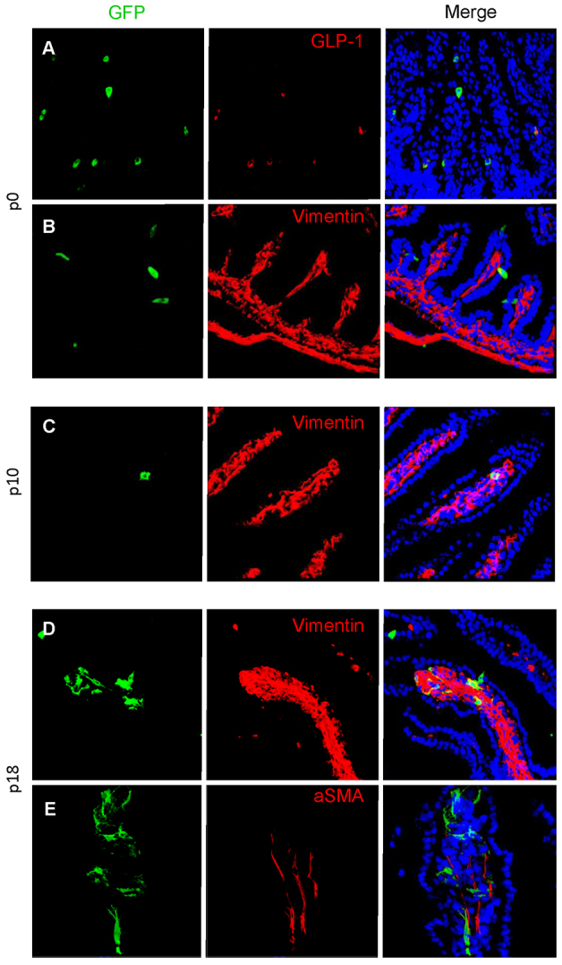
Evidence for epithelial-to-mesenchymal transition. At P0, GLU-Cre/ROSA26-GCaMP3 mice have GFP expression limited to epithelial cells within the small intestine, shown by cells colocalising with GLP-1 (A). No mesenchymal cells were observed within the mesenchymal core of villi (n=3 animals), although some epithelial GFP cells colocalise with the mesenchymal marker vimentin (B). In P10 mice, GFP cells are present that do not colocalise with proglucagon but have a mesenchymal appearance and co-stain with the mesenchymal marker vimentin (C). Finally, in P18 mice, larger numbers of GFP-expressing mesenchymal cells are present. A subset of these co-stain with vimentin (D), whereas others co-stain with SMA (E).