
Fig. 2.
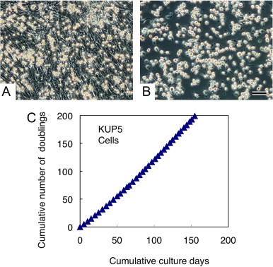
Selective isolation of immortalized C57BL/6 mouse Kupffer cells by a shaking and attachment method. Kupffer cells that proliferated on the mixed cell sheet (A) were infected with a retroviral vector containing the human c-myc oncogene and neomycin resistance gene. After infection for 3 days, Kupffer cells were suspended into the culture medium by gently shaking the flasks, subsequently transferred into a non-tissue culture grade plastic dish and incubated for 30 min at 37 °C. Kupffer cells promptly attached to the dish surface, while other, contaminating fibroblastic cells remained suspended. After a rinse with PBS, a highly purified Kupffer cell population was obtained (B). After selection and cloning with neomycin containing medium, the KUP5 cell line was established. The stable proliferative capacity of KUP5 cells was demonstrated by the continuous passage at 4–5 days intervals for 5 months with a constant population doubling time of approximately 19 h (C). Scale bar = 100 µm.