Figure 2.

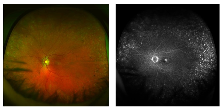
Optomap colour and fluorescein angiogram images showing extensive drusen, RPE mottling, and small areas of RPE atrophy in the retinal periphery in an eye with retinal angiomatous proliferation.

Optomap colour and fluorescein angiogram images showing extensive drusen, RPE mottling, and small areas of RPE atrophy in the retinal periphery in an eye with retinal angiomatous proliferation.