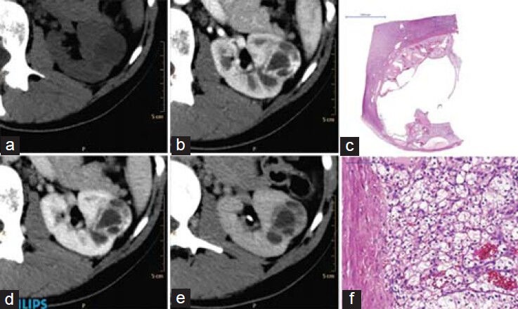
Figure 1.

Computed tomography (CT) appearance of a typical Bosniak III category cystic lesion. Thickened irregular wall and septas with contrast enhancement can be observed in the lesion located in the middle third of the left kidney. Unenhanced (a), corticomedullary (b), nephrographic (c) and excretory phases (d) axial CT images. Histological H and E stained sections displaying an intracystic clear cell renal cell cancer at magnification ×1 (e) and ×20 (f)
