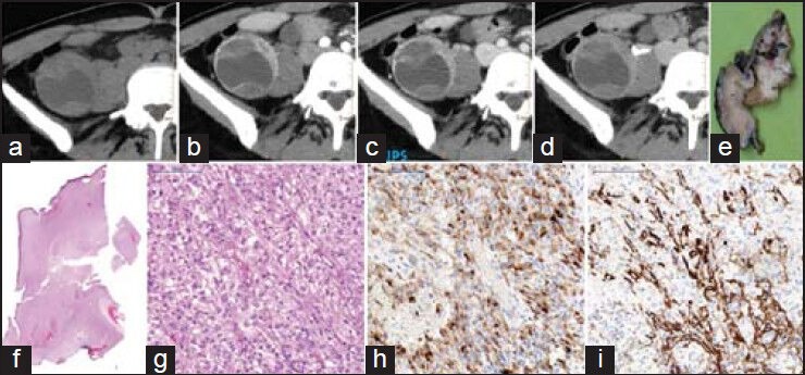
Figure 4.

Computed tomography (CT) appearance of the atypical angiomyolipoma mimicking a Bosniak category III lesion. 37-year-old asymptomatic lady. Thickened, irregular wall and septa with contrast enhancement can be appreciated in the lower third of the right kidney. Unenhanced (a), corticomedullary (b), nephrographic (c) and excretory phases (d) axial CT images. Macroscopic documentation (e) with H and E stained histological sections at magnification ×1 (f) and ×20 (g). Immunohistochemical images of HMB-45 (h) and SMA (i) of the identical region of the tumor at magnification ×20
