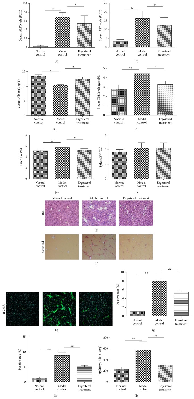
Figure 4.
Antifibrotic effects of ergosterol on CCl4-induced liver fibrosis in mice. Mice of ten-week old were treated as described in the legend. Serum ALT (a), AST (b), Alb (c), and T.Bil (d) were assessed by liver function tests kits, respectively. Levels of serum liver functions were decreased, and ratios of liver/BW (e) and spleen/BW (f) were alleviated by Ergosterol treatment in CCl4-induced liver fibrosis mice. Histologic results of liver tissues were stained with H&E ((g), ×200). Quantification of the same three groups in (g) with respect to Sirius Red staining ((h), ×100). Collagen deposition was shown as percentage of Sirius red-positive area (j). Liver tissues were analyzed by staining for α-SMA immunofluorescence. Representative bright-field and fluorescent micrographs were shown ((i), ×100). Semiquantification data for α-SMA expressions in the liver tissue were evaluated and were shown as percentage of α-SMA-positive areas (k). Hydroxyproline content was quantified from 100 mg liver samples and measured by Jamall's method (l). ** P < 0.01, versus Normal control; ## P < 0.01, versus Model control.

