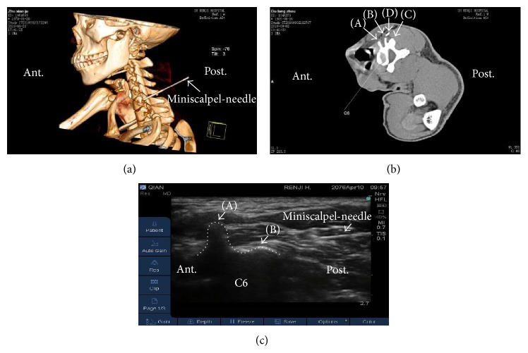
Figure 3.
UG-MSN release at the sixth cervical vertebra (C6). (a) Three-dimensional computed tomography reconstruction of the puncture site at C6. The white arrow indicates the MSN. (b) Conventional computed tomography scanning of C6. The letters (A)–(D) indicate, respectively, the anterior tubercle, posterior tubercle, articular process, and the MSN. (c) UG-MSN release at C6, with the MSN clearly indicated. Trigger points showed the following characteristics: hyperechoic skin, adipose tissue of mixed echogenicity, and muscle of hyperechoic, marbled appearance. The dotted line shows the outline of C6. The letters (A) and (B) represent, respectively, the posterior tubercle and articular process. Post.: posterior position of patients. Ant.: anterior position of patients.

