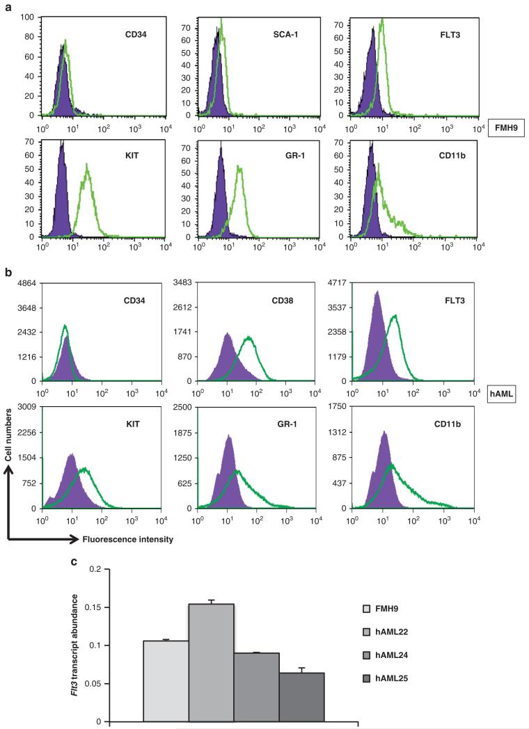
Figure 1. FMH9 cells and AML cells flow cytometric characterisation.
(a) (b) Staining with labelled antibodies against stem cell-related antigens (KIT, SCA-1, CD34, FLT3) and lineage specific markers (GR-1, CD11b) are overlaid against matched isotype controls. (c) FLT3 mRNA quantification by quantitative-PCR, normalised against the 18S house keeping gene results.

