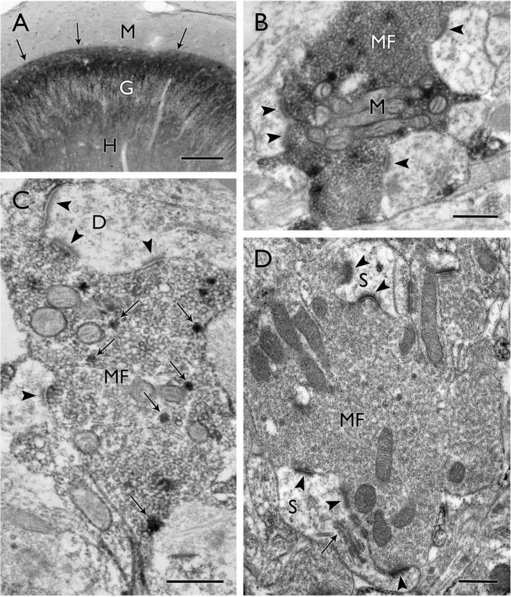
Figure 1.

Mossy fiber reorganization in surgical specimens from humans with temporal lobe epilepsy. (A) Dynorphin-labeled mossy fibers form a distinct, aberrant band (arrows) in the inner molecular layer (M), immediately above the granule cell layer (G), while the normally strong dynorphin labeling in the hilus (H) is reduced. (B) A large dynorphin-labeled mossy fiber terminal (MF) contains a cluster of mitochondrial profiles (M) and forms asymmetric synaptic contacts (arrowheads) on at least three different dendritic profiles in this single electron micrograph. (C) In a more lightly-labeled mossy fiber terminal (MF), dynorphin-labeled dense core vesicles (examples at arrows) are scattered among the numerous clear synaptic vesicles that fill the terminal. The terminal forms multiple asymmetric synaptic contacts (arrowheads) on one dendritic profile (D) and an additional synaptic contact on another. (D) A large mossy fiber terminal (MF) is packed with synaptic vesicles and forms synaptic contacts with complex spines (S). In the upper part of the field, two distinct synaptic contacts (arrowheads) are separated by a protrusion from a spine, creating a perforated synapse. In the lower part of the field, multiple synaptic contacts (arrowheads) are formed with a large spine that contains a spine apparatus (arrow). Scale bars = A, 200 μm; B–D, 0.5 μm.
