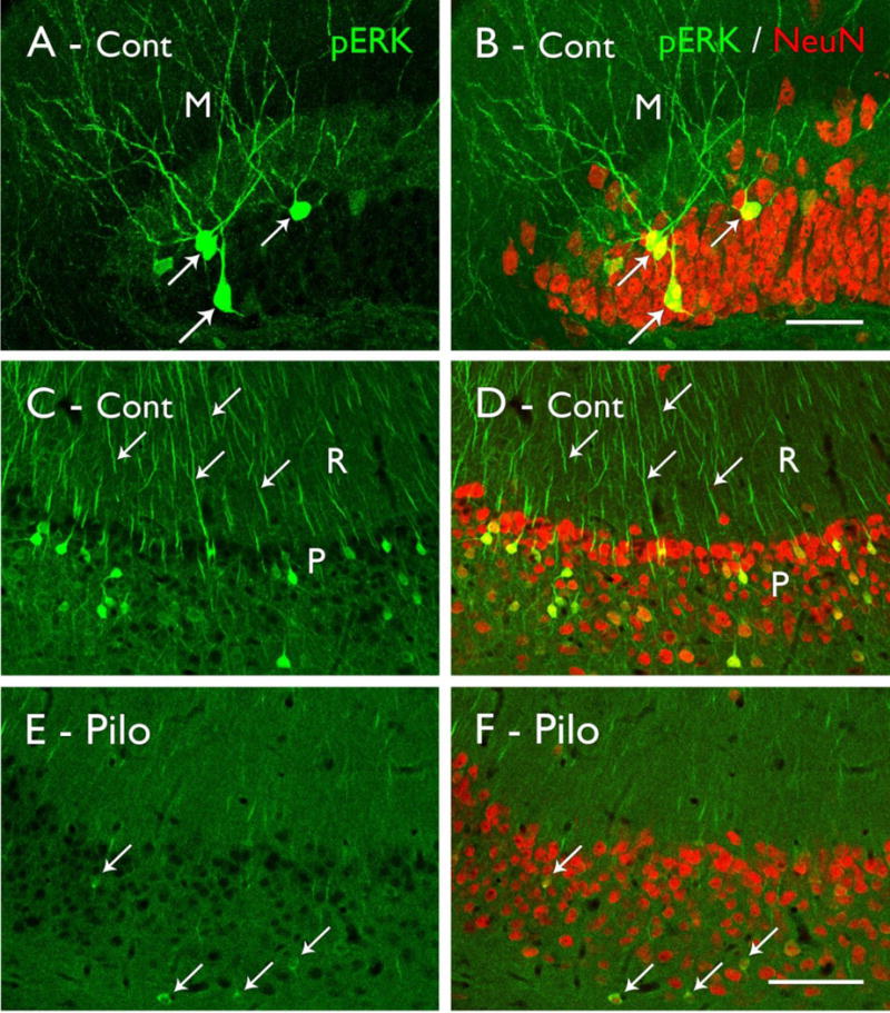
Figure 3.

Phosphorylated ERK (pERK) labeling in control (Cont; A–D) and pilocarpine-treated (Pilo; E,F) mice. (A,B) Strong pERK labeling is evident in scattered granule cells in the dentate gyrus (arrows), and their labeled dendrites extend into the molecular layer (M). Double labeling with NeuN demonstrates the relatively small proportion of granule cells that are strongly labeled for pERK in control animals. (C,D) Strong pERK labeling is also evident in selected neurons in the pyramidal cell layer (P) of CA1, and their apical dendrites (examples at arrows) extend into s. radiatum (R). Double labeling with NeuN demonstrates that pERK-labeled neurons are distributed throughout the pyramidal cell layer. (E,F) In a pilocarpine-treated mouse, the numbers of neurons that are strongly labeled for pERK are substantially reduced, although faint labeling can be detected in some neurons (arrows). Double-labeling with NeuN demonstrates that the reduced labeling is not due to cell loss in the pyramidal cell layer in this animal. (Scale bars = A,B, 20 μm; C–F, 100 μm.
