Figure 4.

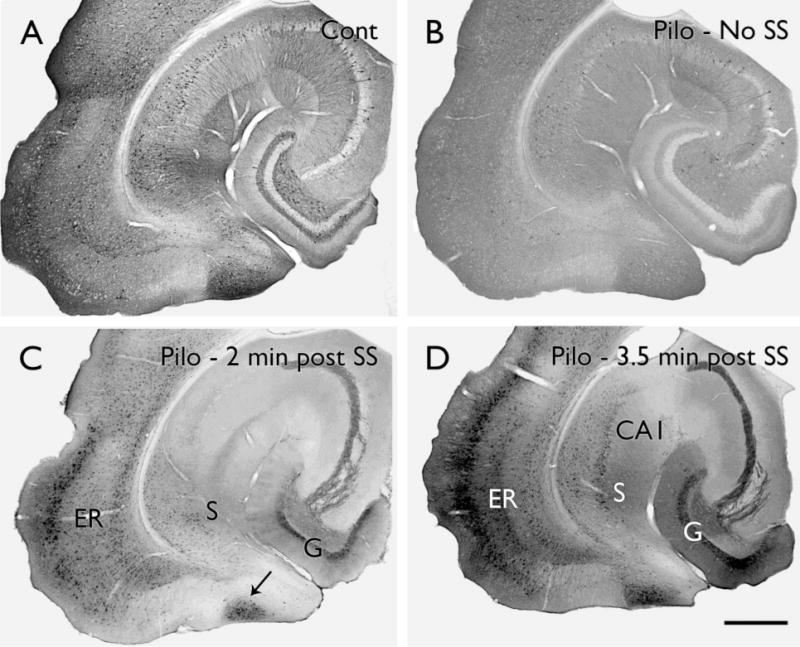
Alterations in phosphorylated ERK labeling in pilocarpine-treated mice. (A) In a control (Cont) mouse, scattered, strongly-labeled neurons are evident in most brain regions and stand out against a background of diffuse pERK labeling. (B) In a pilocarpine (Pilo)-treated mouse that had not experienced a spontaneous seizure (SS) during the previous 24 hours, the number of neurons with strong pERK labeling is substantially decreased throughout much of the hippocampal formation and associated limbic cortex. (C) In a pilocarpine-treated mouse at approximately 2 minutes after detection of a spontaneous behavioral seizure, pERK labeling is strongly increased in several limbic regions, including a large extent of the dentate granule cell layer (G), lateral entorhinal cortex (ER), subiculum (S) and a unique zone (arrow) that is located between the parasubiculum and medial entorhinal cortex. (D) In a mouse at 3.5 minutes after a spontaneous seizure, pERK labeling is substantially increased and is strong throughout the granule cell layer, lateral entorhinal cortex, CA1 and subiculum. Scale bar = A–D, 200 μm.
