Figure 3.
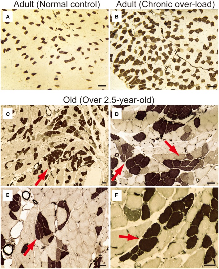
ATPase staining for adult (A, 15-week-old) and old-aged (C–F, 136–139-week-old) rat PLT muscles. ATPase staining was performed after incubation at pH 4.3; thus, Type I fibers were stained dark. Note that (B) is a comparative positive control for ATPase staining using compensatory hypertrophied PLT muscle after 6 weeks of surgical ablation of synergistic gastrocnemius and soleus muscles (16-week-old rats). This demonstrated that the shift to slow fibers occurred after chronic stretch stimulation. However, slow-type fiber grouping was evident in the old-aged PLT muscle (C), suggesting remodeling of S-type motor units in the process of sarcopenia (red arrows in C–F). Panels (D–F) shows higher magnification of (C). Bars in (A,B) = 100 μm, (C–F) = 50 μm.

