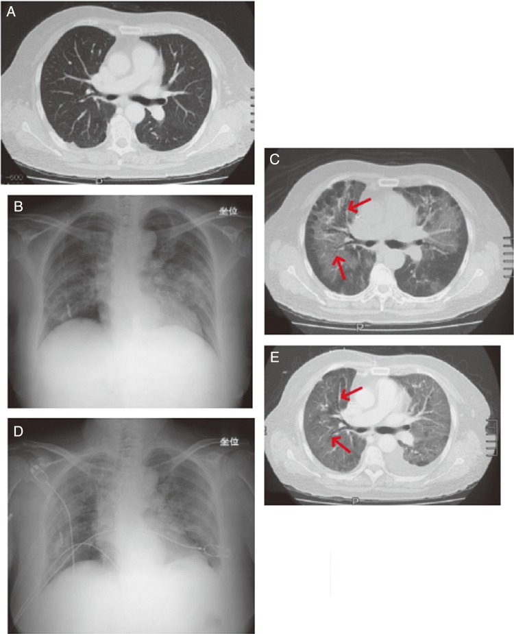
Figure 3.
Case report of a female patient with recurrent colorectal cancer in her 60s. (A) Image before the onset of DLI taken at 32 days after starting cetuximab therapy. Metastatic nodes were observed in the inferior lobe of both lungs. An infiltrate and ground-glass opacity were not observed. (B) X-ray image taken 60 days after starting cetuximab therapy, 3 days after the onset of symptoms. Ground-glass opacity was predominantly observed in the bilateral upper lung field. (C) Computed tomography (CT) scan on Day 60. Ground-glass opacity was observed from the hilus to the middle section of both lungs. The lesion in the periphery of the lung field was lightly spared. Hypertransradiancy of bronchi (arrow) was observed, but there was no obvious traction bronchiectasis, and it could not be diagnosed as diffuse alveolar damage from the images. (D) X-ray image taken on Day 67. The abnormal shadows in both lung fields had spread and were exacerbated. (E) CT image taken on Day 68. The shadows in both lungs were exacerbated and extended to the periphery of each lung. Hypertransradiancy of bronchi with bellows-like dilation (arrow) was observed in the shadows, and was considered to be traction bronchiectasis. The organizing stage of diffuse alveolar damage was observed, which was considered to be a sign of lesion progression. Left pleural effusion was also observed.

