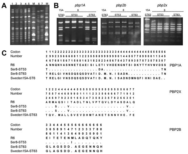
Figure 2.

A) Pulsed-field gel electrophoresis patterns of chromosomal DNA of Streptococcus pneumoniae isolates after digestion with SmaI. Lane 1, Sweden 15A-ST63 (American Type Culture Collection [ATCC] BAA-661); lanes 2−5, serotype 8-ST63; lanes 6 and 7, serotype 8-ST53; lane M, molecular mass markers. B) PCR–restriction fragment length polymorphism patterns of penicillin-binding protein genes pbp1A, pbp2b, and pbp2x. Lane 1, Sweden 15A-ST63 (ATCC BAA-661) lanes 2−4: serotype 8-ST63; lanes 5 and 6, serotype 8-ST53. C) Amino acid sequence variations among PBP1A, PBP2B, and PBP2X proteins. Row 1, S. pneumoniae R6; row 2, serotype 8-ST53; row 3, serotype 8-ST63; row 4, Sweden 15A-ST63 reference strain. Codon numbers of polymorphic sites are numbered in a vertical format. Amino acids are numbered according to their positions in the corresponding protein. Only those amino acids that differ from those of the strain R6 sequence are shown. Dots indicate an amino acid residue that is identical with that in the strain R6 sequence.
